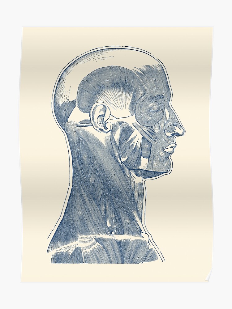
Head And Neck Muscle Diagram - Superficial Muscles of the Face and Neck : The deep neck muscles (figure 7.18 and table 7.7) include neck flexors, located along the anterior surfaces of the vertebral bodies, and neck extensors, located posteriorly.
Head And Neck Muscle Diagram - Superficial Muscles of the Face and Neck : The deep neck muscles (figure 7.18 and table 7.7) include neck flexors, located along the anterior surfaces of the vertebral bodies, and neck extensors, located posteriorly.. Click here to read about mesothelioma and its differential diagnosis and mesothelioma t. For more anatomy content please follow us and visit our website: Masster, temporalis, medial pterygoid and lateral pterygoid muscles. They move the head in every direction, pulling the skull and jaw towards the shoulders, spine, and scapula. They move the head in every direction pulling the skull and jaw towards the shoulders spine and.
Instant anatomy is a specialised web site for you to learn all about human anatomy of the body with diagrams the muscles of the neck anatomical chart shows in beautiful detail the many anterior, posterior, inferior and lateral views of every muscle that makes. Deep muscles of the neck. The deep neck muscles (figure 7.18 and table 7.7) include neck flexors, located along the anterior surfaces of the vertebral bodies, and neck extensors, located posteriorly. Left versus right brain traits diagram. This article describes the anatomy of the head and neck of the human body, including the brain, bones, muscles, blood vessels, nerves, glands, nose, mouth, teeth, tongue, and throat.
This muscle originates at some of the spinous processes in your thoracic vertebrae (the thoracic vertebrae are the ones where your ribs connect), and it inserts at the transverse processes of the first.
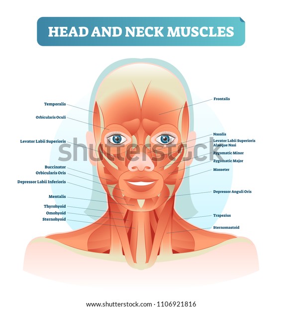
Muscles of the neck typically have actions associated with movement of the head and swallowing. Zenon yeung captured excellent photographs during dissections and miss janet fong crafted the professional diagrams from our. These muscles are located medially to the ears, superior to the mandible, and inferior to the coronal suture of the skull. Human anatomy diagrams show internal organs, cells, systems, conditions, symptoms and sickness information and/or tips for healthy living. Angled muscles from the cheekbones to the corners of the mouth ( origin zygomatic bone, action elevate corners of the mouth. The head and neck is characterized by its complex anatomy that performs vital functions and is dr. They move the head in every direction pulling the skull and jaw towards the shoulders spine and. Only two of the more obvious and superficial neck. This article describes the anatomy of the head and neck of the human body, including the brain, bones, muscles, blood vessels, nerves, glands, nose, mouth, teeth, tongue, and throat. Start studying 08 muscles of head and neck. Left versus right brain traits diagram. Deep muscles of the neck. Mouse over each are to identify the muscle.
Learn this topic fast with head and neck muscle anatomy reference charts. Body spine bifid spinous process carotid tuber. Angled muscles from the cheekbones to the corners of the mouth ( origin zygomatic bone, action elevate corners of the mouth. Muscles of the neck typically have actions associated with movement of the head and swallowing. The head and neck is characterized by its complex anatomy that performs vital functions and is dr.
This muscle originates at some of the spinous processes in your thoracic vertebrae (the thoracic vertebrae are the ones where your ribs connect), and it inserts at the transverse processes of the first.
The quizzes below each include 15 multiple choice identification questions related to the muscles of the head and neck. Body spine bifid spinous process carotid tuber. Masster, temporalis, medial pterygoid and lateral pterygoid muscles. 9 видео 363 062 просмотра обновлен 5 февр. Key facts about head anatomy. Angled muscles from the cheekbones to the corners of the mouth ( origin zygomatic bone, action elevate corners of the mouth. Rotation and lateral flexion of the head are accomplished by lateral and posterior neck muscles. Mouse over each are to identify the muscle. This muscle originates at some of the spinous processes in your thoracic vertebrae (the thoracic vertebrae are the ones where your ribs connect), and it inserts at the transverse processes of the first. Start studying 08 muscles of head and neck. Zenon yeung captured excellent photographs during dissections and miss janet fong crafted the professional diagrams from our. We hope this picture head and neck muscles diagram can help you study and research. These muscles are located medially to the ears, superior to the mandible, and inferior to the coronal suture of the skull.
Key facts about head anatomy. Head and neck surgery the chinese university of hong kong. They move the head in every direction, pulling the skull and jaw towards the shoulders, spine, and scapula. The deep neck muscles (figure 7.18 and table 7.7) include neck flexors, located along the anterior surfaces of the vertebral bodies, and neck extensors, located posteriorly. 9 видео 363 062 просмотра обновлен 5 февр.

Human muscle system, the muscles of the human body that work the skeletal system, that are under voluntary control, and that are concerned with movement, posture, and balance.
To know the answers, please click on the text highlighted in blue. For more anatomy content please follow us and visit our website: Instant anatomy is a specialised web site for you to learn all about human anatomy of the body with diagrams the muscles of the neck anatomical chart shows in beautiful detail the many anterior, posterior, inferior and lateral views of every muscle that makes. Orbital, nasal, oral, auricular and scalp/neck muscles. Eukaryotic vs prokaryotic cells, educational biology vector illustration diagram. The head rests on the top part of the vertebral column, with the skull joining at c1. Masster, temporalis, medial pterygoid and lateral pterygoid muscles. Body spine bifid spinous process carotid tuber. Learn this topic fast with head and neck muscle anatomy reference charts. Head and neck surgery the chinese university of hong kong. Human muscle system, the muscles of the human body that work the skeletal system, that are under voluntary control, and that are concerned with movement, posture, and balance. Human anatomy diagrams show internal organs, cells, systems, conditions, symptoms and sickness information and/or tips for healthy living. Free download abdomen,spleen,liver anatomy and physiology diagrams.
Zenon yeung captured excellent photographs during dissections and miss janet fong crafted the professional diagrams from our neck muscle diagram. Eukaryotic vs prokaryotic cells, educational biology vector illustration diagram.